A collection of inspiring news from SCUBA Diving Community
Our aim is to provide a compelling narrative and case for change to inspire people all over the world.
In Medicine
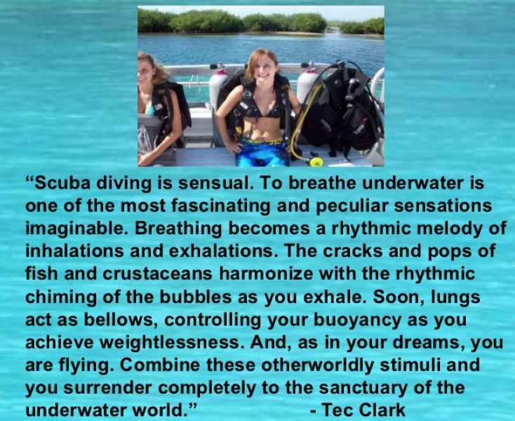
For years, I thought the episode was pretty bogus. Even as a kid I knew that becoming a scuba diver required hours of arduous training. I never gave the show much thought until years later when, following in my hero’s footsteps, I, too, became a scuba instructor. Even back then, everyone wanted to be like Mike. I soon came to realize that my hero hadn’t let me down after all. In fact, his 30-second lesson was brilliant in its cut-to-the-chase elegance, and certainly could have been enough training given the dire circumstance and high motivation of the unlucky victim. I don’t remember the dialogue verbatim, but it went something like this: “You see this thing? [pointing to the regulator mouthpiece] You put it in your mouth and breathe. Whatever you do, keep breathing; don’t ever hold your breath, or your lungs will burst and you’ll die!”
It was one of the few instances where Hollywood actually got the facts straight. If you had but 30 seconds to teach someone to scuba dive, what would you tell them? The same thing Mike did — the Golden Rule of scuba diving. Breathe normally; never hold your breath. The rest, in most cases, is pretty much secondary.
Of course, if you’re learning to dive without the distraction of gunfire, and your instructor has a bit more time to explain the nuances and importance of breathing, you probably will be subjected to either an illustration or an actual example of the most commonly used prop in diver training — the ubiquitous balloon. And the explanation, though lacking the dramatic effect that Lloyd Bridges could bring to the lesson, will be something like: If a flexible, gas-filled container — like a lung — can’t vent excess pressure as it rises in the water column, its volume will expand until it bursts. Of course, today you might have sophisticated video or computer-based graphics, but the essence of what Mike told the scientist remains the same.
Unfortunately, the balloon-aided explanation is about all that most divers ever learn. Now, there’s nothing really wrong with the balloon analogy. It’s just a bit oversimplified, especially if you really want to fully understand the consequences of forgetting what Mike Nelson so succinctly told the scientist. For one thing, our lungs bear little resemblance to balloons. (A sponge is a much more accurate analogy.) And due to the intricate and delicate nature of their anatomy, severe problems occur from lung expansion long before, as Mike so aptly put it, “your lungs burst and you die.”
Human lungs are amazing structures, made up of microscopically small air sacs called alveoli. While incredibly tiny, the massive number of alveoli — numbering in the hundreds of millions — provides an enormous surface area for gas exchange. How large, you ask? If the total surface area of the alveoli were somehow laid flat, it would cover an area two-thirds the size of a tennis court!
And talk about delicate — alveoli redefine the term. Their walls are only one cell thick. Yet as small as they are, each alveolus (that’s singular for alveoli, for those of you whose native tongue isn’t Latin) is surrounded by numerous capillaries that take up oxygen and give off carbon dioxide. And how about this for amazing: These capillaries are so small that red blood cells pass through in single file. Equally incredible is that while the distance between the gas in the alveoli and the blood in the capillary is less than one-twentieth the thickness of this page, at no time is the blood in the capillaries directly exposed to air.
The fact that alveoli are so delicate may be interesting, but it’s also at the root of the problem when it comes to lung expansion injuries. Unlike the highly elastic balloon you probably heard about in your Open Water course, the alveoli of the human lungs can’t quite take the same licking and keep on ticking. In fact, in shallow water, alveolar rupture can occur with an increase in internal pressure of a mere 70 mmHg (millimeters of mercury) — about 2 psi — over the external pressure. Why’s that important? It means that if you hold a full breath in shallow water, an ascent of only 4 feet (a little over a meter) could ruin your day.
This brings up the first of many interesting contrasts between lung expansion injuries and decompression sickness (DCS). Unlike DCS, where the risk increases with depth, lung expansion problems are more likely in shallow water (above 2 atmospheres) because of the greater change in volume. (To understand this, think back to the trusty old balloon. If brought from 33 feet/10 m to the surface, it will show a 100-percent increase in volume. But bringing the same balloon from 66 feet/20 m to 33 feet will result in only a 50-percent volume increase, and only a 33-percent increase from 99 feet/30 m to 66 feet). One should need no more rationale than this to explain to your untrained friends why it’s not safe to “play around” with your tanks even in the seemingly innocuous environment of a swimming pool.
Lung expansion injuries can be divided into at least three types, depending on where the bubbles go once they leave the alveolus. The most critical injury happens when air escapes directly into the tiny capillaries surrounding the lung, and the mechanism involved is very interesting. First, over-pressurization of the alveolus forces air into the surrounding blood vessels. This often results from a tear in the alveolar wall. But believe it or not, because of the tiny size and delicate structure of the alveoli, in some cases gas can escape without actually tearing lung tissue.
Regardless of how the air gets out of the alveoli, it can’t begin its trip to the brain just yet.
At first, expansion of the overstretched alveoli compresses the blood vessels, preventing air bubbles from entering the bloodstream. Only when the alveolar expansion is relieved through exhalation can the air bubbles make their way into circulation. Only a very small amount of air is required to produce ill effects. It’s estimated, for example, that gas bubbles as small as 30 microns — that’s 30 millionths of a millimeter — are sufficient to cause symptoms.
Once in the capillaries, air bubbles flow into the pulmonary veins, which lead to the heart. From there they merge on to the superhighway of the body’s blood vessels and enter the arterial circulation. Like the tiny balloon your instructor told you about, the bubbles continue to expand as the ascent progresses. And as the bubbles continue on their journey, the circulatory system branches into smaller and smaller vessels. At some point the bubbles become larger than the diameter of the vessel containing them, and a blockage occurs. Such a blockage is called an embolus, and in this case is caused by air or gas.
Furthermore, in divers the condition normally occurs in the brain. (Another example of an embolus would be dislodged plaque from an artery that causes a stroke.) Thus, the term in medical parlance for this condition is cerebral arterial gas embolism, or CAGE.
As no more blood can flow to affected regions due to the blockage, the tissues served by the vessel are starved for oxygen. The brain is the body’s control center, and as the blockage can occur virtually anywhere in the brain, the signs and symptoms of air embolism are highly variable. In most cases, however, the onset of symptoms is sudden (within one minute of surfacing), dramatic and may appear stroke-like. Specifically, victims may experience: dizziness, blurred vision, disorientation, personality change or decreased level of consciousness, paralysis or weakness, bloody froth from the mouth or nose, convulsions, shock, unconsciousness and respiratory arrest.
Some version of what was just explained is about all that most divers know about lung expansion. But there’s more. For example, the gas escaping from an alveolar rupture doesn’t necessarily have to enter the circulatory system. Instead, the bubbles can escape and lodge between the lung tissue and capillaries, and track along the loose tissue plates surrounding the airways and blood vessels. From there the gas may continue to travel into the mediastinum (the medical term for the space containing all the organs of the chest except the lungs), into the region surrounding the heart (pericardium) or up to the base of the neck. In the case of the air in the mediastinum, the condition is called a pneumomediastinum or mediastinal emphysema. Involvement of the pericardium is called pneumopericardium, and air under the region of the neck is dubbed subcutaneous emphysema.
In severe cases symptoms of these disorders may appear immediately, but in less severe cases it can take several hours. For pneumomediastinum or mediastinal emphysema, the symptoms may include: chest pain (usually under the breastbone), breathing difficulty or discomfort, fainting, shock or a change in voice. In subcutaneous emphysema, symptoms are: swelling or feeling of fullness around the neck, significant voice change, difficulty swallowing and a crackling sensation when the skin is pressed. The formal medical term for this is crepitus.
Finally, the air may escape by rupturing the visceral pleura — the inner layer of the membrane surrounding the lungs — and enter the pleural cavity. This condition is called a pneumothorax (literally, “air in the chest”). As there is no tissue connecting the lungs to the chest wall, any air introduced into the pleural cavity causes the lung to collapse. If the air space does not expand, the condition is called a simple pneumothorax. If the air space does expand, it puts tension on the heart and interferes with breathing and circulation.
This condition is known as a tension pneumothorax and can result from expansion of the air space in the chest on ascent, or even after the diver has surfaced with a simple pneumothorax. A tension pneumothorax is a very severe problem requiring immediate medical intervention. Symptoms of both types of pneumothorax include: sudden unilateral chest pain with movement, breathing difficulty or very rapid breathing rate, and blue skin, lips or nailbeds.
You, as well as your instructor, have probably always assumed that lung expansion problems happen only to divers who hold their breath because of some distraction, lack of training or panic situation. In some instances, air can remain trapped in the lungs even when the diver exhales. Some of the factors that can promote air trapping include: asthma, bronchitis, cysts, tumors, scar tissue from surgery or radiation therapy, and obstructions from inflammation or mucus caused by smoking, and even recent colds or infection. This is why candidates for diving must be free of any serious pulmonary disease or other chronic problems involving the lungs. For this reason, some medical opinions advocate chest x-rays and other pulmonary tests for diving candidates. In fact, in Australia, full medical exams, including chest x-rays, are required for anyone who enrolls in a scuba course. This is not the case here in North America, although some diving physicians feel it should be.
Because both conditions are caused by bubbles, divers are often confused by the differences between air embolism and decompression sickness. On the surface, the distinction seems clear: An air embolism is a traumatic injury resulting from a mechanical rupture of the alveoli which introduces air bubbles into the arterial circulation. DCS is caused by supersaturating of inert gas and results when dissolved nitrogen comes out of solution to form gas bubbles. In fact, to emphasize their distinct causation, instructors often place great stress on the differences between air embolism and DCS.
More recently, however, this delineation has become blurred, and has caused many to rethink how divers should be trained and how medical authorities should describe the disorders. In fact, it’s now believed that while they’re caused in fundamentally different ways, the disorders nonetheless could have significant interaction.
For example, some physiologists believe that many unexplained cases of DCS actually begin as subtle air embolism events. The mechanism works as follows: What could be termed a “micro air embolism” occurs, introducing tiny amounts of air into the bloodstream. Then, the high level of supersaturated nitrogen begins dissolving into the bubbles, causing them to grow. Without the air bubbles to act as “seeds” for further bubble growth, DCS would probably not have occurred. But is such an event a case of air embolism or DCS? The answer is, probably a little of both. So in an attempt to merge these complicated, interrelated and speculative processes, scientists and clinicians now use the term decompression illness (DCI) to identify what have classically been described separately as air embolism and decompression sickness.
Perhaps the most important practical reason for downplaying the differences between air embolism and DCS in diver training has to do with first aid measures. While the differences between the two disorders may be interesting in an academic sense, from the standpoint of how you assist an injured diver, such differences are irrelevant. Anyone suspected of having decompression illness — regardless of whether the condition is caused by mechanical injury or supersaturation — is treated the same way at the scene of an accident. (See sidebar on first aid measures.)
One aspect of managing a diving casualty that still evokes some confusion, and even controversy, is the issue of head position. For many years divers were taught to use a left-side-down, head-low position for treating diving accident victims. Most often this was accomplished by putting the entire body on an incline (called the Trendelenburg Position). The idea was that in such a position, the buoyant force of the bubbles would resist moving in a downward path, thus reducing the tendency for bubbles to travel toward the brain or heart and make the condition worse.
But practical experience has shown the T-position to be of no use to victims of DCS, and to have little, if any, positive effect on most air embolism victims. Moreover, in some cases it has actually caused complications, such as breathing difficulty and edema of the brain. Without a backboard, placing and keeping a victim in such a position is very difficult. So a few years ago the guidelines changed to simply keeping the victim in a left-side-down position, with no head tilt or incline.
Still, there are those who believe that a slight head-low position could be beneficial for a victim who is likely suffering from air embolism. Advocates point to both animal studies and anecdotal experience to support their opinion. Currently, the advice from medical authorities is that if a head-low position is used, it should only be done for a short period (20 minutes or less), and only if it does not impair breathing or interfere with other first aid measures such as CPR. Under no circumstances, whether the victim is in a head-low position or merely lying flat, should he or she be allowed to raise the head.
Although air embolism commonly accompanies the various other forms of emphysema and pneumothorax, that’s not always the case. But as embolism is the most serious disorder, it’s important that it’s ruled out even in cases where only the other disorders are suspected. These are, of course, medical decisions that can be made only by a qualified physician.
Cases involving emphysema only are most often managed solely by observation and having the patient breathe pure oxygen. Treatment for pneumothorax alone depends on the severity of injury. Minor cases (less than 20-percent collapse) may require nothing more than medical observation. In more severe cases, the air must be extracted surgically. In either case, recompression is rarely used.
Air embolism is a completely different matter. Immediate recompression is absolutely essential, and treatment has historically involved compressing the victim in a chamber to 6 atmospheres, an equivalent depth of 165 feet/50 m. (This is far deeper than the maximum depth of 60 feet used to treat DCS.) The extreme pressure reduces gas bubbles to one-sixth of their surface volume. This helps restore blood flow and promotes reabsorption of the bubbles. More recently, however, some clinicians have revised this treatment procedure, and first take the patient down to 60 feet for observation. If he responds, the descent is halted and treatment continued at that depth. (One reason this method is beneficial is that many chambers don’t have the capability of going to 6 atmospheres.)
The ironic part about lung expansion problems is that while they are the most serious injuries divers face, they’re also the easiest to prevent. Accident data also has provided two important lessons. First, as one might expect, lung expansion injuries occur primarily — though not exclusively — in novices and less-experienced divers. Second is some good news: Recent accident analyses show the frequency of such injuries is declining (accounting for only 90 of 935 diving injuries in 1996). This is partly attributed to the increased awareness of the value of slow ascent in preventing DCS and the increased popularity of dive computers (which all have ascent rate monitors, often with audible alarms). I wonder what Mike Nelson would have to say about all this?
Like decompression sickness, one can never completely eliminate the risk of a lung expansion injury. But you can reduce your chances of injury. Here’s how:
1. Ascend slowly. Even if you’re breathing normally, a rapid ascent rate could lead to a lung overpressure injury through gas trapping.
2. Use a high-quality regulator and have it serviced regularly. It’s believed by some that excessive inhalation effort may cause edema (fluid damage) to tissues surrounding the alveoli, thus reducing the size and impeding flow into and out of the airway.
3. Avoid diving too soon after a chest cold or respiratory infection. This means that no matter how good you feel, don’t dive if you are coughing up mucus, or if your breathing produces any abnormal noise or resistance. To reduce the tendency for mucus obstruction after a chest cold, drink plenty of water before diving.
4. Running out of air is the major cause of lung expansion problems, so practice good air management techniques. Have enough air to make the dive you’re planning — plus some reserve. Monitor your own and your buddy’s gauges frequently.
5. Forget what you were told about a 60-foot-per-minute ascent rate being OK. Slow down to half that. It will help you avoid both lung expansion injuries and DCS.
6. Don’t smoke, and if you do, stop. Smoking causes the buildup of mucus, which can obstruct airways.
While the differences in symptomology between DCS and lung overpressure injuries can be subtle, this is of no concern to divers at the scene of an accident. Regardless of which disorder is actually present (sometimes both are), the first aid measures are the same. The information below was excerpted from DAN’s Underwater Diving Accident and Oxygen First Aid Manual.
1. Administer CPR if required, with victim lying flat (supine).
2. Keep airway open and prevent aspiration of vomitus. Unconscious victims should be intubated by trained personnel.
3. Administer oxygen by tight-fitting, transparent, double-seal mask at the highest possible oxygen concentration. Do not remove oxygen except to reopen the airway or if victim shows signs of convulsions.
4. Keep victim in the horizontal left-side-down position if symptoms occurred within 10 minutes of surfacing and steps 1 through 3 have been completed.
5. If convulsion occurs, do not forcefully restrain. Turn victim on side (supporting head and neck), maintain airway and sweep away any vomitus. Hold diver loosely to prevent self-injury, and do not forcefully insert an airway or tongue blade. Resume oxygen administration when convulsions cease.
6. Protect the victim from excessive heat, cold, wetness or noxious fumes.
7. For Conscious Victims Only — Give nonalcoholic liquids orally, such as water or fruit juices,
8. Transport the diver to the nearest emergency room to be evaluated and stabilized in preparation for transport to a recompression chamber.
9. Call DAN at (919) 684-8111. State that you have an emergency, and ask for the person on call. (If necessary, call collect in an emergency.)
10. If air evacuation will be used, it is critical that the victim not be further injured by exposure to decreased barometric pressure at altitude. Flight crews must maintain cabin pressure at sea level or fly at the lowest safe altitude in unpressurized aircraft.
11. Contact hyperbaric trauma center or chamber before transporting the victim.
12. If available, send a copy of DAN’s Underwater Diving Accident and Oxygen First Aid Manual, and record history (dive profile, diver’s complaints, medical history and first aid) of the victim.
13. Send all diving equipment with the victim for examination. If that’s not possible, arrange for local examination and gas analysis.
As a child of the ’50s, I was a big fan of the hallmark TV series Sea Hunt and its indomitable hero, Mike Nelson, played by the late actor Lloyd Bridges. One episode I particularly remember involved the kidnapping of a scientist. As this was the era of Sputnik, it was implied — though never overtly stated — that the culprits were a group of “stinking commies.” The scientist was being held in a cave on an island. Central to the story line was the fact — seemingly unknown to the bad guys — that the cave could be entered from underwater. Of course, Mike Nelson knew all about the underwater entryway and planned a highly sophisticated escape: he swam into the cave, distracted the guards, gave the scientist a 30-second scuba lesson, dodged a few bullets on the way out and — fade to black — the world was once again a safe place for mom, apple pie and clean-cut capitalists.
Visit Page